 OptiWave Refractive Analysis (ORA)
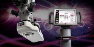
OptiWave™ Refractive Analysis, ORA for short, is a revolutionary new, cutting edge technology that can be used during your cataract procedure to optimize your postoperative visual outcomes. ORA provides an on-demand analysis of your eye during surgery, not possible with today’s conventional measurements and instruments. At any point in the cataract procedure, your surgeon can easily take a measurement, which is then analyzed and used to guide your surgeon’s decision making to optimize the vision of your eye.
ORA works by directing a beam of low intensity laser light into the eye during the surgical procedure. The laser light reflects off the back of your eye, and sensors in the ORA device analyze the reflected wave of light exiting your eye. This real-time analysis measures all of the eye’s unique optical characteristics, and gives your surgeon an accurate measurement of your eye’s focusing capabilities.
Click here to download a list of frequently asked questions about ORA.
Ocular Coherence Tomography (OCT)
Optical coherence tomography (OCT) is a new technology that provides noninvasive, high resolution, cross-sectional imaging of biological tissues. The test can image retinal structures in the eye with a resolution of 10 to 17 microns. The anatomic layers within the retina can be differentiated and retinal thickness can be measured. This exciting new technology also has the potential to revolutionize the early detection of glaucoma through its ability to evaluate the nerve cells damaged in glaucoma. Of particular clinical importance at the present time is the early and accurate detection and staging of macular holes, often a severe form of vision loss and in the localization of fluid accumulation within the retina, such as can be found in central serous retinopathy or diabetic maculopathy. OCT can assist in identifying a variety of ocular pathologies including diabetic retinopathy, macular holes, epiretinal membranes, cystoid macular edema, central serous choroidopathy and optic disc pits.\
A-Scan
A-scan ultrasound biometry , commonly referred to as an A-scan , is a routine test performed to measure the shape of the eye before cataract surgery. A-scan measurements determine the axial eye length which is used by the surgeon in the calculation of intraocular lens power during the pre-operative assessment for cataract surgery.
Fundus Photos
Fundus photos are simply pictures taken with a specialized camera that reveal the interior posterior surface (back) of the eye, including the retina, optic disc, macula and posterior pole. Your physician will order these photos to rule out any disease processes that might be affecting these structures.
Fluorescein Angiography
Fluorescein angiography is a test commonly ordered by a retinal specialist. The test is used for evaluating retinal, choroidal and iris blood vessels, as well as any eye problems affecting these vessels. To perform the test, a dye is injected into an arm vein, after which a series of sequential photographs are taken of the internal eye as the dye circulates. The contrast of the dye going through the veins allows the retinal specialist to assess any potential disease states. Your physician will discuss potential risks associated with the test.
Visual Field Test
The visual field test assesses the potential loss of peripheral vision and maps the loss to assist the physician with determining the cause. The test requires a patient to fix on a central target point and note when objects appear in their peripheral vision. Visual fields are often used to follow progress of glaucoma patients. Other diseases that may affect the visual field of the eye include diabetes, stroke, hyperthyroidism, hypertension, diseases of the pituitary gland, and multiple sclerosis.


